The electron microprobe

The ultimate gemological instrument?
The electron microprobe is also called an EPMA, which stands for electron probe microanalyzer. The microprobe is similar to a scanning electron microscope (SEM) in that it uses an electron beam in a vacuum chamber, but rather than imaging, its primary purpose is to provide a chemical analysis of a very small volume of solid material.
Microprobes are most often used by hard rock geologists (e.g. mineralogists and igneous and 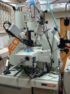 metamorphic petrologists), paleontologists and sedimentologists as well as materials scientists and are generally only found at major universities. The instruments have been commercially available since the 1960s and new instruments currently cost in excess of $1 million.
Compared to other pieces of analytical instrumentation the microprobe has some unique characteristics:
-
1)Unlike most quantitative techniques, the analytical method is non-destructive which makes it particularly suitable for the analysis of gemstones.
-
2)Sample preparation requires a relatively small (< 40mm x 20mm) polished specimen, or gem, but is relatively simple and straightforward. Very tiny fragments or grains can be analyzed.
-
3)Full quantitative chemical analysis is possible with high accuracy for major, minor and trace elements. Detection limits are typically 100 ppm but this is misleading in that compared to highly sensitive bulk analytical techniques like instrumental neutron activation analysis a microprobe can detect an amount of material one hundred thousand times smaller.
-
4)Elements from atomic number 3 through 92 can be analyzed.
-
5)Analysis time per specimen is generally quite short (<5 min) and throughput is quite good.
-
6)Amazing spatial resolution on the order of one to two micrometers (10-6 m) {a micrometer is about 1/25th the diameter of the human hair} allows for studies of zoning, textural analysis, diffusion and inclusions, etc.
Geologists generally rely on polarized light microscopy to identify rocks and minerals as crushed powders or thin sections. The microprobe provides a full chemical analysis of the specimen typically providing an unambiguous identification. In most cases the chemistry is enough to identify the mineral, but in some cases the structure of the mineral needs also to be defined, in which case X-ray diffraction techniques are implemented requiring either single-crystal or powder diffractometers.
METHOD
EPMA uses a focused high-energy beam of electrons in a vacuum chamber. The electron beam current is typically 10-200 nanoamps, approximately 1000 times stronger than that used in an SEM. When the beam strikes the target, x-rays are emitted that are characteristic of the elements present and the x-rays are diffracted by analyzing crystals and counted by gas-flow or proportional detectors. The chemistry of the sample is then determined by comparison to standards of known chemical composition.
In practice, once inside the probe, the chemistry of the sample is first qualitatively assessed by an energy dispersive x-ray spectrometer that using a solid state detector rapidly determines the elements present in rough relative amounts displayed in graphical form. The operator then knows what elements to look for and tunes the wavelength dispersive spectrometers to detect those elements and derive the quantitative data.
I have recently purchased an electron microprobe and photos of the instrument and its supporting equipment can be found in the the Blog section of this website.
APPLICATIONS TO GEMOLOGY
Competent gemologists can identify the vast majority of gem species using the common gemological instruments, but occasionally there is a vexing I.D. problem that can only be solved by chemical, spectral or structural analysis. In addition, gem treatments or synthetics can generally be detected by variations in chemistry. Likewise, the country or location of origin can often be determined by trace element chemistry or the identification of characteristic inclusions.
Today, most of the major inclusions in gemstones have been identified and documented, mainly by Gubelin and Koivula in their extraordinary three volume set, Photoatlas of Inclusions in Gemstones. Methods of practical inclusion I.D. include:
1) site identification from experience with inclusions and the common associations of inclusions with gemstone provinance
-
2)using an optical goniometer to determine crystal face angles in euhedral crystals
-
3)use of the Raman microscope, a relatively recent technique
-
4)collecting micro-powders from exposed inclusions for x-ray powder diffractometry
-
5)exposing inclusions at the surface of the crystal or gem and using the EPMA.
In the early days of inclusion research the electron microprobe was the definitive means of inclusion analysis for serious investigators and it is still the ultimate tool for determinative inclusion I.D. In the author’s opinion, the EPMA is the most important analytical instrument for gem research, but by far the least used because of its expense and the paucity of skilled operators and accessible instruments. One could literally do 100 years of relevant gem research with a microprobe.
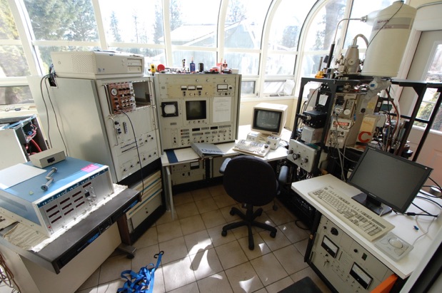
My electron microprobe on the sunporch of my house. Support equipment includes, gaseous high pressure nitrogen, liquid nitrogen in a 25 liter cryogenic dewar, an air compressor, two rotary vacuum pumps and a diffusion pump, a water cooling system, 36000 BTU of air conditioning, a 240V rotary phase converter and the equipment seen here. The copious supply of liquor required to keep from committing suicide from frustration is not shown.